Colon IHC whole-slide quantification
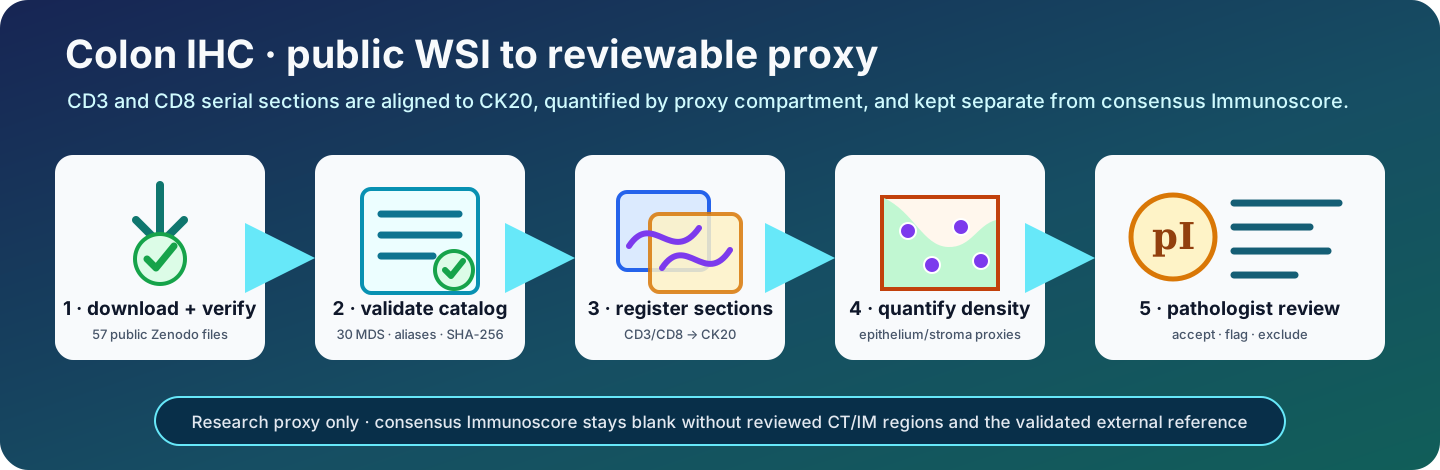
Published reference dataset
The exact reference release is public and requires no Zenodo credential:
| Field | Published value |
|---|---|
| Dataset | TumorQuantAI colon cancer CD3, CD8, and CK20 whole-slide image dataset |
| Record | 22177196 |
| DOI | 10.5281/zenodo.22177196 |
| Version | 1.0.0 |
| Files | 57 public files; 40,721,516,620 bytes |
| WSIs | 30 sanitized MDS files: 10 CD3, 10 CD8, and 10 CK20 |
| Cases | 11 aliases; 9 complete triplets and 2 explicitly unavailable incomplete cases |
| Rights statement | Copyright (C) 2026 The Authors; public visibility does not itself create a separate open-content license |
The deposit includes the MDS files, exact SHA-256 and MD5 rosters, a case/slide/marker catalog, the complete TumorQuantAI value and QC CSVs, nine registration composites, 36 paper-ready review sheets, an HTML report, and an offline pathologist accept/flag/exclude dashboard. The dataset DOI identifies this dataset—not the TumorQuantAI software and not a clinically validated Immunoscore assay.
Research proxy—not clinical Immunoscore
This workflow does not calculate the clinically validated consensus
Immunoscore. The required pathologist-reviewed tumour core (CT), invasive
margin (IM), and validated external reference population are absent.
TumorQuantAI therefore leaves consensus_immunoscore blank and reports its
status as unavailable. It also emits an explicitly provisional pI0–pI4
within-cohort analogue for pathologist accept/flag review. The four density
measurements, internal percentiles, and pI label are research outputs only.
What this workflow answers
Why this is not the consensus assay
The validated colon-cancer Immunoscore measures CD3 and CD8 cell densities in the tumour core and invasive margin. The invasive margin is a 720 µm region centred on the tumour boundary, and four density percentiles are referenced to a validated 700-case population before they are averaged. Pathologist review of the tumour and margin regions is part of that process.
This dataset instead contains adjacent CD3, CD8, and CK20 sections. CK20 helps separate epithelial from non-epithelial tissue, but it does not establish CT and IM. CK20 expression is linked to epithelial differentiation and can vary between the tumour centre and leading edge, so it cannot define the entire invasive boundary. TumorQuantAI makes the distinction explicit:
| Output | Region definition | Reference distribution | Intended use |
|---|---|---|---|
| Consensus Immunoscore | Pathologist-validated CT and 720 µm IM | Validated external 700-case cohort | Not available here |
| TumorQuantAI CK20-guided proxy | CK20-positive epithelial proxy and CK20-negative common-tissue proxy | This cohort only | Research exploration and QC |
| TumorQuantAI provisional pI0-pI4 | Mean of four CK20-proxy percentiles | Automatic-QC-pass cases in this run | Prioritize pathologist accept/flag review only |
See the consensus validation in The Lancet, the analytical validation protocol, the CK20 spatial-expression caveat, and the VALIS serial-WSI registration paper.
Install
git clone https://github.com/cfarkas/tumorquantai.git
cd tumorquantai

./tumorquantai install --conda
export PATH="$HOME/.local/bin:$PATH"

tumorquantai --inmunoscore --help
The command is package-native and does not invoke HistoPLUS or Nextflow.
tumorquantai ihc immunoscore is the canonical subcommand;
tumorquantai --inmunoscore is the requested top-level compatibility form.
Reproduce the public analysis
The public route consumes the flat Zenodo MDS filenames and the published
tumorquantai_colon_immunoscore_slide_catalog.csv directly. It preserves the
published TQA_CI_... case aliases, requires no alias secret, and creates no
private linkage table.
1. Download all 57 files
The release is 40.7 GB. Keep it outside the Git checkout and allow enough additional space for a fresh result directory. The following resumable command uses the Zenodo API only to obtain the fixed filenames and public content URLs:
TQA_PUBLIC="$HOME/tumorquantai-data/colon-ihc-v1.0.0"
install -d "$TQA_PUBLIC"

curl -fsSL --retry 5 https://zenodo.org/api/records/22177196 \
  | jq -r '.files[] | "\(.links.self)\n  out=\(.key)"' \
  | aria2c --input-file=- \
      --dir="$TQA_PUBLIC" \
      --continue=true \
      --max-concurrent-downloads=3 \
      --max-connection-per-server=1 \
      --auto-file-renaming=false \
      --allow-overwrite=true
The command was tested against the published endpoint and preserves each
Zenodo filename. Rerunning it resumes partial transfers. jq and aria2c must
be installed by the operating system; neither is a TumorQuantAI runtime
dependency.
2. Verify the immutable payload
Verify the primary SHA-256 roster before opening or analysing the images:
(cd "$TQA_PUBLIC" && sha256sum --check SHA256SUMS)

find "$TQA_PUBLIC" -maxdepth 1 -type f -name 'TQA_CIS_*.mds' | wc -l
# Expected: 30
SHA256SUMS covers the other 55 release files; the two checksum rosters do not
hash themselves. MD5SUMS is supplied for repository interoperability and can
be checked separately with md5sum --check MD5SUMS. SHA-256 is the primary
integrity check.
3. Preview the public catalog
TQA_RESULTS="$HOME/tumorquantai-results/colon-ihc-v1.0.0-reproduction"

tumorquantai --inmunoscore "$TQA_PUBLIC" \
  --output "$TQA_RESULTS" \
  --public-slide-catalog \
    "$TQA_PUBLIC/tumorquantai_colon_immunoscore_slide_catalog.csv" \
  --workers 3 \
  --dry-run
For the published catalog, the preview must report:
input_identity_mode: published_public_slide_catalog
discovered_slide_count: 30
discovered_case_count: 11
complete_case_count: 9
incomplete_case_count: 2
marker_slide_counts: CD3=10, CD8=10, CK20=10
source_mpp_values: [0.26178]
The fail-closed reader checks the exact catalog columns, alias shapes, one
slide per case/marker, safe flat filenames, byte sizes, physical scale, source
format, sanitization profile, and equality between the catalog and input MDS
rosters. --dry-run does not hash image payloads or create output.
4. Quantify from the public MDS files
After the preview matches, remove only --dry-run:
tumorquantai --inmunoscore "$TQA_PUBLIC" \
  --output "$TQA_RESULTS" \
  --public-slide-catalog \
    "$TQA_PUBLIC/tumorquantai_colon_immunoscore_slide_catalog.csv" \
  --workers 3
Before case analysis, TumorQuantAI hashes every catalogued MDS and requires its
size and SHA-256 to match the publication catalog. The run is resumable and
writes new results outside the downloaded release. Do not point --output at
the Zenodo directory.
Review the published output
You do not need to rerun 30 WSIs to inspect the reference predictions.
Download the complete review-figure ZIP (108 MiB) Open the case-value CSV
Direct public result downloads:
- paper-ready case and marker figures (ZIP, 108 MiB);
- TumorQuantAI case values (CSV);
- cohort density summary (CSV);
- cohort report (HTML);
- pathologist review dashboard (HTML); and
- all 57 files on the Zenodo record.
Zenodo deliberately serves the HTML files as plain text. If clicking an HTML link shows source code, use the browser's Save link as action. Save the report and dashboard to the same local directory as the extracted figure bundle, then open them locally. To use the local web-server option, expand the ZIP beside the two HTML files and keep this command running in its terminal:
(cd "$TQA_PUBLIC" && unzip -q -o tumorquantai_immunoscore_paper_figures.zip)
(cd "$TQA_PUBLIC" && python3 -m http.server 8000 --bind 127.0.0.1)
Only then, on that same computer, open
http://127.0.0.1:8000/REPORT.html for the frozen cohort report and
http://127.0.0.1:8000/PATHOLOGIST_REVIEW.html for case adjudication.
127.0.0.1 is not a public TumorQuantAI address: it always means the computer
running the browser. If the command runs on a remote SSH server, download the
bundle locally or use an SSH port forward; do not expose the review server to
the public Internet.
The dashboard displays the case sheet from the extracted cases/ tree, falls
back to the deposited registration composite when necessary, preserves all
TumorQuantAI fields, and exports pathologist_review_completed.csv with
additive accept, flag, or exclude decisions.
Treat completed review exports as controlled research records
The blank deposited dashboard is public, but reviewer codes, free-text notes, and downstream annotations can introduce sensitive or identifying content. Inspect a completed export before sharing it.
Published result snapshot
| Cohort result | Published value |
|---|---|
| Automatic-QC pass | 6 cases |
| Automatic-QC review | 3 cases |
| Unavailable incomplete marker set | 2 cases |
| Provisional pI distribution | pI0: 3; pI1: 0; pI2: 4; pI3: 2; pI4: 0; unscored: 2 |
| Consensus Immunoscore | 0 reported; blank with explicit unavailable status for all 11 cases |
The nine numerically available cases have these deposited CK20-proxy density summaries:
| Measurement | Median positive cells/mm² | Range |
|---|---|---|
| CD3, CK20-epithelium proxy | 86.46 | 11.25–3,363.76 |
| CD3, CK20-stroma proxy | 107.85 | 22.02–411.68 |
| CD8, CK20-epithelium proxy | 58.86 | 5.57–3,986.58 |
| CD8, CK20-stroma proxy | 68.42 | 6.14–152.66 |
These are descriptive values from the published
cohort_density_summary.csv,
not estimates of clinical performance. The pI distribution is cohort-relative
and must be interpreted only after reviewing registration, CK20 compartment
assignment, stain quality, tissue coverage, and cell segmentation.
Analyze private source bundles
Keep original archives, extracted scanner bundles, alias secrets, and linkage tables outside Git:
controlled-colon-ihc/
├── archives/                         # original ZIPs
├── private_source/
│   └── extracted/inmunoscore/
│       └── <private-case>-CD3-.../
│           ├── 1.mds
│           └── info.ini
├── private_release/
│   ├── alias_secret.bin              # mode 0600, at least 32 random bytes
│   └── case_slide_linkage.csv         # created by the run; mode 0600
└── results/
Create the one-time secret without printing it:
install -d -m 700 /controlled/colon-ihc/private_release
head -c 32 /dev/urandom > /controlled/colon-ihc/private_release/alias_secret.bin
chmod 600 /controlled/colon-ihc/private_release/alias_secret.bin
Do not regenerate this file between runs. The same secret and source IDs produce the same aliases; another secret produces unrelated aliases.
How cases are linked
TumorQuantAI does not infer case identity from staining intensity, filenames that merely look similar, or pathologist values. It uses an exact rule:
- The immediate bundle name must match
<source-case-id>-CD3|CD8|CK20-<scanner-suffix>. - The exact text before the marker token is the private case ID.
- HMAC-SHA-256 with the owner-controlled secret creates one
TQA_CI_...case alias. - The complete bundle name independently creates a
TQA_CIS_...slide alias. - A source case may have at most one slide for each marker.
The controlled CSV records the complete evidence chain:
case_alias
slide_alias
source_case_id
source_slide_id
marker
source_mds_path
source_mds_size_bytes
source_mds_sha256
Only case_alias, slide_alias, marker, and non-identifying acquisition facts
appear in public results. The private linkage is required for exact audit and
must never be uploaded to Zenodo or committed to Git.
Run a private cohort
Set paths once:
TQA_MDS=/controlled/colon-ihc/private_source/extracted/inmunoscore
TQA_RESULTS=/controlled/colon-ihc/results/tumorquantai_immunoscore
TQA_SECRET=/controlled/colon-ihc/private_release/alias_secret.bin
TQA_LINKAGE=/controlled/colon-ihc/private_release/case_slide_linkage.csv
1. Inventory-only preview
tumorquantai --inmunoscore "$TQA_MDS" \
  --output "$TQA_RESULTS" \
  --alias-secret-file "$TQA_SECRET" \
  --private-linkage "$TQA_LINKAGE" \
  --workers 3 \
  --dry-run
The preview reads the private Motic info.ini scale and reports complete and
incomplete marker sets. It does not hash WSIs, create linkage, or write output.
2. Quantify
Remove only --dry-run:
tumorquantai --inmunoscore "$TQA_MDS" \
  --output "$TQA_RESULTS" \
  --alias-secret-file "$TQA_SECRET" \
  --private-linkage "$TQA_LINKAGE" \
  --workers 3
The run is resumable by default. It verifies the exact private linkage before
reusing a completed case and refuses a non-empty foreign output directory.
Use --no-resume only when intentionally starting with a new empty output.
What TumorQuantAI does
1. Read the Motic pyramid safely
The package opens only DSI0 pixel tile streams from each MDS compound file.
Label, macro, and acquisition streams are inaccessible to the quantifier.
Physical scale comes from the private Motic info.ini; an optional
--source-mpp assertion must agree with it exactly.
The analysis level nearest 0.55 µm/pixel is selected by default. For scans at 0.261780 µm/pixel this is the 0.5 pyramid level, 0.523560 µm/pixel.
2. Register serial sections
Bounded overviews are formed from DSI0 tiles. SIFT and ORB feature candidates,
partial/full affine transforms, and a tissue-bounding-box fallback are scored
by tissue Dice. The best geometrically valid transform is retained. A Dice
below --minimum-registration-dice never becomes a silent zero; it produces a
review status. The feature-free tissue-bounding-box fallback is also always
review-only, even when its global tissue Dice is high.
3. Form CK20-guided proxy compartments
Direct stain separation on a 2048-pixel WSI overview can average focal brown glands into the background. TumorQuantAI therefore streams the CK20 pyramid at the level nearest 2.0 µm/pixel (2.094240 µm/pixel for this cohort), performs colour-checked H–DAB separation in each block, and area-projects the fraction of expected-brown pixels into the bounded registration overview. An overview pixel enters the raw proxy when at least 2% of its contributing fine pixels pass the 0.08 DAB-OD threshold.
Connected regions smaller than 1,000 µm² are removed, subject to a four-pixel
minimum at the realised overview resolution. Retained regions are closed and
expanded by 8 µm to form ck20_epithelium_proxy; common registered tissue
outside the mask is ck20_stroma_proxy. Pyramid coordinates are converted by
their numeric level scale and the explicit overview resize—not by padded tile
canvas ratios. This matters because MDS levels can have different amounts of
white edge padding.
Every measurement.json records the selected CK20 level, realised µm/pixel,
block counts, fine-pixel DAB fraction, projected-fraction threshold and range,
raw/final compartment fractions, overview scale, and effective morphology.
Treat these assignments as coarse CK20-guided WSI compartments, not
cellular-resolution tumour or invasive-margin annotations.
4. Stream CD3/CD8 object detection
Each immune WSI is processed in bounded blocks, so the full 100,000-pixel-scale image is never materialized in memory. Counterstained and DAB-positive objects are watershed-segmented. A 3 µm expanded-cell region is positive when either:
mean expected-brown DAB OD ≥ 0.16
OR
at least 8% of expanded-cell pixels have DAB OD ≥ 0.16
An 8 µm strip is excluded at block boundaries from cell centres and the corresponding analysed footprint to reduce clipped/duplicate objects. A marker with no segmented nuclei remains numerically auditable but is forced to review status; it cannot enter the pass-only cohort statistics or ranks.
5. Map and quantify
Every retained centroid is transformed from the immune analysis level through the moving overview into CK20 coordinates. This transform uses the numeric MDS pyramid scale plus any explicit overview resize so differing tile padding does not shift cells. Counts are divided by the matching common-tissue compartment area:
positive-cell density = mapped positive cells / analysed compartment area (mm²)
The four headline TumorQuantAI values are:
tumorquantai_cd3_ck20_epithelium_density_per_mm2
tumorquantai_cd3_ck20_stroma_density_per_mm2
tumorquantai_cd8_ck20_epithelium_density_per_mm2
tumorquantai_cd8_ck20_stroma_density_per_mm2
6. Generate a provisional score without impersonating validation
Automatic-QC-pass cases define four deterministic mid-rank reference distributions. Every numerically available pass or review case is placed against those references. TumorQuantAI averages its four percentiles and emits:
pI0   0–10
pI1  >10–25
pI2  >25–70
pI3  >70–95
pI4  >95–100
The bands mirror the published five-category percentile presentation, but the
mandatory p prefix marks a different, provisional calculation: CK20
epithelium/stroma substitutes for CT/IM, and this small internal cohort
substitutes for the validated external distribution. Review cases can receive a
pI value so the pathologist can accept or flag the technical result, but they
never contribute to the reference. consensus_immunoscore remains blank.
7. Build paper-ready review sheets
TumorQuantAI writes a 300-dpi case summary plus one PNG/PDF/legend triplet for each CK20, CD3, and CD8 WSI. The layout follows the useful conventions of WSI plotters such as LazySlide: clean overview panels, explicit overlays, physical scale bars, and export-resolution metadata. It adds the exact four density values, a pI gauge, and QC flags required for this workflow. See LazySlide's WSI visualization guide and publication-quality export guidance for the visual conventions; TumorQuantAI's renderer is independent and adds no LazySlide runtime dependency.
The immune panels are registration blends at overview scale. They are not cell-outline overlays. Open the source WSI at cellular resolution whenever segmentation itself is in question.
Review a new local run
Open:
results/tumorquantai_immunoscore/START_HERE.html
Review in this order:
- Confirm the expected complete and incomplete marker sets.
- Open every
registration_qc.png. - Reject or correct implausible serial-section registration.
- Review the CK20 epithelial proxy across both tumour centre and leading edge.
- Check analysed area and mapped-positive-cell fraction.
- Open
PATHOLOGIST_REVIEW.html, inspect the 300-dpi paper sheet, and chooseaccept,flag, orexclude. - Export
pathologist_review_completed.csv; preserve the original prediction and automatic-QC columns unchanged. - Only then interpret the reviewed density and provisional-score tables.
The output map is:
tumorquantai_immunoscore/
├── START_HERE.html
├── PATHOLOGIST_REVIEW.html
├── cases/<case_alias>/
│   ├── measurement.json
│   ├── registration_qc.png
│   └── paper_figures/
│       ├── case_summary_paper_figure.{png,pdf}
│       ├── case_summary_paper_figure_legend.txt
│       └── <slide_alias>_<marker>_paper_figure.{png,pdf}
├── tables/
│   ├── public_slide_inventory.csv
│   ├── tumorquantai_immunoscore_values.csv
│   ├── cohort_density_summary.csv
│   ├── case_compartment_densities.csv
│   ├── registration_qc.csv
│   ├── paper_figure_manifest.csv
│   ├── pathologist_review_template.csv
│   ├── pathologist_review_codebook.csv
│   └── unavailable_cases.csv
└── workflow_metadata/
    ├── immunoscore_run.json
    └── failures.json                    # only when a case failed
What the numbers mean
| File | Use it for | Important boundary |
|---|---|---|
tumorquantai_immunoscore_values.csv |
One row per public case alias, four densities, internal percentiles, pI0-pI4, and explicit consensus-unavailable fields | Review cases are scored against—but never included in—the automatic-pass reference |
cohort_density_summary.csv |
Mean, sample SD, median, quartiles, and range for each density under pass-only and all-numeric populations | Population and n are explicit; review cases never silently enter pass-only statistics |
case_compartment_densities.csv |
Counts, areas, densities, mapping fraction, MPP, and QC in long format | A technically completed row can still require review |
registration_qc.csv |
Transform method, feature matches, inliers, tissue Dice, and affine matrix | Automated Dice does not replace visual review |
paper_figure_manifest.csv |
Every 300-dpi case/slide PNG, PDF, legend, and layout version | Overview-scale figures do not prove cell-level segmentation accuracy |
pathologist_review_template.csv |
Blank accept/flag/exclude adjudication fields beside immutable algorithm outputs | Exported review must retain reviewer code and timestamp; it never overwrites predictions |
unavailable_cases.csv |
Missing-marker and failed-case audit | Missing is never encoded as biological zero |
Zenodo privacy boundary
The release workflow creates new MDS files under non-semantic HMAC-derived slide aliases. It preserves every DSI0 pixel stream byte-for-byte and replaces all label, macro, and non-pixel OLE streams with deterministic same-size generic neutral content. The replacement payload never repeats a source stream name. Each staged file is then checked for:
- exact DSI0 stream names and bytes;
- full and sampled DSI0 fingerprints;
- deterministic non-pixel replacement;
- absence of case and bundle-name variants in UTF-8, Latin-1, UTF-16LE, and UTF-16BE;
- decodability and pyramid geometry;
- visual tissue-pixel review.
The original ZIPs, sidecars, source filenames, alias secret, private linkage,
and private sanitizer mapping remain outside the deposit. The package release
helper can create only a restricted, unsubmitted Zenodo draft; it contains no
publication action. For reference release 1.0.0, the owner separately
completed the privacy, integrity, visual, rights, and metadata gate, published
the record, and explicitly changed all 57 files to public visibility. That
manual action produced DOI
10.5281/zenodo.22177196; it is not
performed by TumorQuantAI.
Non-semantic public aliases and sanitized container metadata do not justify attempting re-identification from tissue pixels or combining the images with outside identifying data.
Limitations to carry into every report
- Serial sections are not cell-for-cell identical.
- CK20 is an epithelial aid, not a complete colorectal tumour-boundary marker.
- The DAB thresholds are deterministic research settings, not assay calibration.
- Automatic tissue Dice can miss local misregistration.
- No tumour core or invasive margin has been reviewed.
- Internal ranks are cohort-relative and will change with cohort composition.
- Outputs must not guide patient care.
For the exact CLI contract and CSV schemas, see CK20-guided CD3/CD8 reference.