Breast IHC quantification
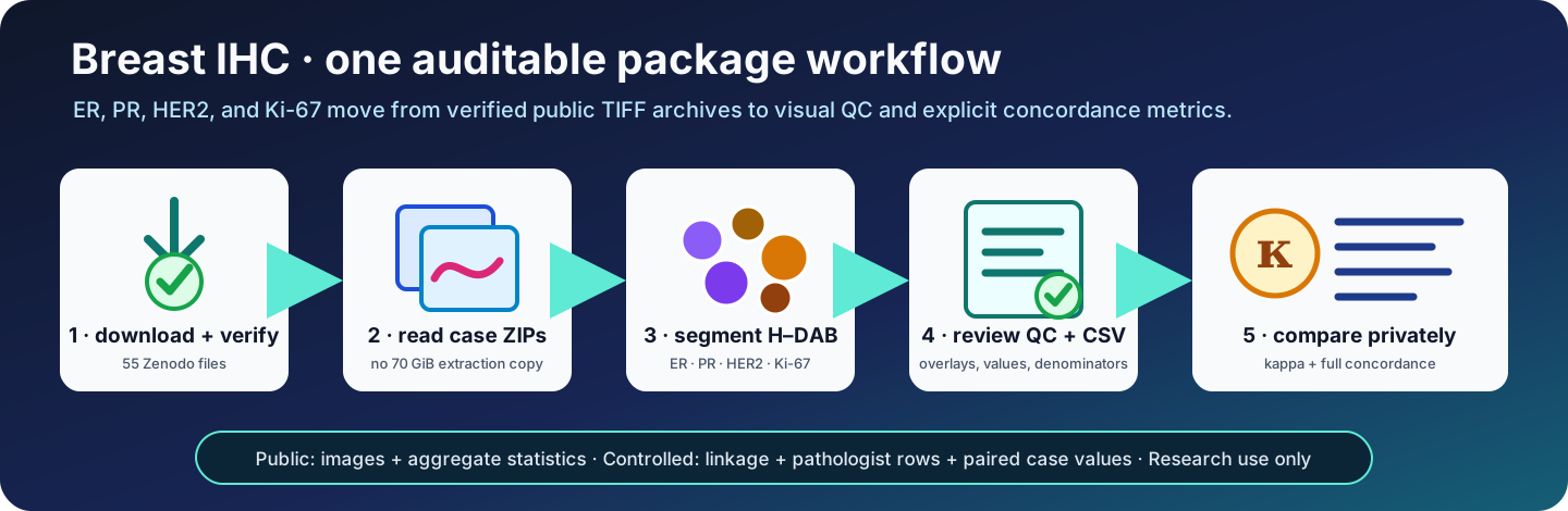
Research use only
TumorQuantAI produces computational research pre-scores. They are not diagnoses, clinical assay results, treatment recommendations, or evidence of clinical validation. The public fields have no pathologist-verified invasive-tumour ROI and no validated tumour-cell classifier.
What this workflow answers
Before you begin
The dataset is public under CC BY 4.0 at
Zenodo record 21797920, DOI
10.5281/zenodo.21797920. Its 55
files total 74,958,557,152 bytes: 51 case ZIPs, one manifest bundle, one
packaging report, and two checksum rosters.
Plan for:
- about 69.8 GiB for the download;
- about 4–5 GiB for overlays, reports, checkpoints, and compressed cell tables
when
--save-cellsis enabled; - a local filesystem with reliable free space;
- no HistoPLUS token or model weights for the package-native IHC route.
The command reads TIFF members from the case ZIPs. You only extract the small manifest bundle, avoiding a second roughly 70 GiB image copy.
Install TumorQuantAI
git clone https://github.com/cfarkas/tumorquantai.git
cd tumorquantai

# Install the global command and the package-native IHC dependencies.
./tumorquantai install --conda
export PATH="$HOME/.local/bin:$PATH"

tumorquantai ihc --help
Use --poetry, --docker, or --singularity instead when that is your
supported installation route. IHC quantification itself runs locally and does
not call the gated HistoPLUS model.
Download and verify the dataset
Download all 55 files from the Zenodo record into one directory without renaming them:
breast-ihc-downloads/
├── TQA_BC_<public-case-alias>.zip          # 51 case archives
├── TQA_BreastIHC_manifest_bundle.zip
├── packaging_report.json
├── SHA256SUMS
└── MD5SUMS
Verify the complete payload, then extract only the manifest bundle:
cd /path/to/breast-ihc-downloads
sha256sum --check SHA256SUMS

mkdir -p manifest
unzip TQA_BreastIHC_manifest_bundle.zip -d manifest
Every line must report OK. A checksum proves file integrity; it does not make
the data or method clinically suitable.
Run the cohort
Set paths once so the preview and full run are identical:
TQA_DOWNLOADS=/path/to/breast-ihc-downloads
TQA_MANIFEST="$TQA_DOWNLOADS/manifest/patch_manifest.csv"
TQA_RESULTS=/path/to/breast-ihc-results
1. Preview
The dry run checks the cohort, marker selection, archives, and analysis settings without decoding images:
tumorquantai ihc quantify "$TQA_DOWNLOADS" \
  --manifest "$TQA_MANIFEST" \
  --output "$TQA_RESULTS" \
  --workers 12 \
  --save-cells \
  --dry-run
2. Quantify
Remove only --dry-run:
tumorquantai ihc quantify "$TQA_DOWNLOADS" \
  --manifest "$TQA_MANIFEST" \
  --output "$TQA_RESULTS" \
  --workers 12 \
  --save-cells
The run is resumable. Each completed patch has an analysis-signature-matched
checkpoint, so repeating the same command after interruption reuses valid
work. TumorQuantAI fails closed when an archive is missing or decoded pixels
do not match the manifest. --allow-missing is reserved for an intentionally
incomplete audit; unavailable markers never become numerical zero.
Why the v2 DAB color check matters
A conventional inverse HED matrix can assign a positive numerical DAB concentration to magenta or almost neutral gray pixels even though they are not brown chromogen. In the original reference run this failure was most visible for ER: all 51 cases crossed 1%, including visibly magenta/gray pathologist-negative fields.
The v2 engine retains a pixel in the scoring DAB channel only when its optical densities follow the expected brown-DAB cone:
ODblue - ODgreen ≥ max(0.02, 0.15 × unconstrained DAB OD)
ODgreen - ODred  ≥ max(0.02, 0.15 × unconstrained DAB OD)
The rule uses image color and the published DAB stain vector only. It does not read pathologist values. The unconstrained HED measurement remains beside the color-checked value in every audit table, so the change is visible rather than silently rewriting the old result.
The public cohort exposed the failure and was also used to audit this correction. Although pathologist values are never inputs to image scoring, the before-and-after concordance on these same cases is method development, not independent validation.
Use --unconstrained-dab-color only to reproduce the earlier unconstrained
behavior in a new output directory. --minimum-dab-color-margin-od and
--minimum-dab-color-ratio are expert sensitivity-analysis controls; changing
either creates a different analysis signature.
What is measured
| Marker | Research measurement | Case pre-score |
|---|---|---|
| ER | Accepted nuclei with color-checked nuclear DAB, intensity classes, and H-score | Color-checked DAB-positive percentage |
| PR | Accepted nuclei with color-checked nuclear DAB, intensity classes, and H-score | Color-checked DAB-positive percentage |
| Ki-67 | Accepted nuclei with color-checked nuclear DAB, intensity classes, and H-score | Color-checked DAB-positive percentage |
| HER2 | Color-checked DAB along an expanded-nucleus boundary proxy | Conservative 0 / 1+ / 2+ / 3+ membrane-proxy category |
The implementation uses the optical-density colour-deconvolution framework of Ruifrok and Johnston. Clinical interpretation context is provided by the ASCO/CAP ER and PR update, CAP HER2 guidance, and the International Ki67 Working Group. Those sources do not validate these computational measurements.
Review the results
Open:
breast-ihc-results/START_HERE.html
The report begins with cohort completion and marker distributions, then links to all 51 case galleries. Review in this order:
- Confirm
1,516 / 1,516selected IHC patches completed. - Confirm all decoded-RGB checks and automated QC checks passed.
- Open every case page and inspect segmentation overlays.
- Check patch-level denominators before using a case aggregate.
- Treat technically complete values as research measurements, not validation.
The core output map is:
breast-ihc-results/
├── START_HERE.html
├── case_reports/<case_alias>.html
├── tables/
│   ├── tumorquantai_marker_values.csv
│   ├── case_marker_measurements.csv
│   ├── patch_measurements.csv
│   └── unavailable_patches.csv
├── patches/<case_alias>/<patch_alias>/
│   ├── measurement.json
│   ├── qc_overlay.png
│   └── cell_measurements.csv.gz
└── workflow_metadata/ihc_run.json
Interpretation boundary
ER, PR, and Ki-67 denominators include all accepted segmented nuclei, not verified invasive tumour cells. HER2 is an expanded-boundary proxy, not a clinical membrane-scoring algorithm. Separate stains are unregistered and cannot establish cell-level co-expression. The color check removes known non-brown optical-density artifacts; it does not supply tumour-cell classification, laboratory controls, or clinical validation.
Compare with pathologist values
This step is optional and private. The public deposit deliberately excludes the mapping between source biopsy identifiers and public case aliases. See case linkage and privacy for the complete identity chain, reference-cohort audit, and file-by-file publication boundary.
Create the minimum English CSV
Use only the reviewed Biopsias finales incluidas worksheet and the exact
private release linkage. For this cohort the workbook-side key is Biopsia
and the linkage-side key is case_id:
install -d -m 700 /private/path/breast-ihc-agreement

tumorquantai ihc anonymize-clinical \
  /private/path/pathologist-review.xlsx \
  --sheet "Biopsias finales incluidas" \
  --linkage /private/path/private-linkage.csv \
  --clinical-id-column "Biopsia" \
  --linkage-id-column case_id \
  --output /private/path/breast-ihc-agreement/pathologist-markers-pseudonymized.csv
If identifiers differ between the two reviewed sources, do not match on marker
values. Create an explicitly reviewed, mode-0600 private crosswalk:
linkage_id,clinical_id
PRIVATE_LINKAGE_IDENTIFIER,PRIVATE_WORKBOOK_IDENTIFIER
Then add:
--identifier-crosswalk /private/path/identifier-crosswalk.csv
The package requires a one-to-one crosswalk and records only its checksum and row count in provenance. The controlled reference audit found 48 exact identifier pairs and three unique one-character discrepancies. TumorQuantAI does not perform fuzzy matching: a custodian must review those discrepancies and record them as explicit crosswalk rows. No marker measurement participated in linkage.
The resulting CSV has exactly six English fields:
case_alias
pathologist_er_percent
pathologist_pr_percent
pathologist_her2_ihc_score
pathologist_her2_fish
pathologist_ki67_percent
Names, national identifiers, biopsy identifiers, dates, age, diagnosis, laterality, specimen type, and grade are excluded.
Pseudonymized is not anonymous
Public aliases plus marker values remain pseudonymized health data. Keep the CSV, workbook, linkage, crosswalk, and paired output outside Git and under controlled access. Only aggregate, non-case-level statistics belong in a public repository.
The custodian's separate direct-ID audit CSV contains
case_alias,release_case_id,pathologist_biopsy_id,identifier_match_method,normalized_hamming_distance.
It explains every case association but must remain mode-0600 and controlled;
it is never a report or GitHub artifact. See the
complete linkage record.
Calculate agreement
tumorquantai ihc compare "$TQA_RESULTS" \
  --pathologist-csv /private/path/breast-ihc-agreement/pathologist-markers-pseudonymized.csv \
  --output /private/path/breast-ihc-agreement/report \
  --bootstrap-iterations 10000 \
  --bootstrap-seed 20260829
Open AGREEMENT_REPORT.html. It includes:
- unweighted Cohen's kappa for ER and PR at 1%;
- quadratic-weighted kappa for HER2 on 0 / 1+ / 2+ / 3+;
- quadratic-weighted kappa for Ki-67 percentage deciles;
- a secondary unweighted Ki-67 view at 20%;
- case-resampled percentile 95% intervals;
- observed and expected agreement plus positive/negative specific agreement;
- MAE, RMSE, median absolute error, mean bias, and 95% limits of agreement;
- Pearson correlation, Spearman correlation, and Lin's concordance correlation coefficient;
- both raters' category margins and every contingency matrix;
- a checked-versus-unconstrained DAB impact analysis for ER and PR;
- automatic warnings when a rater uses only one category.
The clearest machine-readable outputs are:
| CSV | Rows | Contents | Access |
|---|---|---|---|
$TQA_RESULTS/tables/tumorquantai_marker_values.csv |
51 | One row per public case with color-checked TumorQuantAI ER, PR, HER2-proxy, and Ki-67 values, unconstrained HED audit percentages for nuclear markers, segmented-object denominators, H-scores, and QC status | Local result |
report/concordance_metrics.csv |
5 | One aggregate row per prespecified marker scale with the full kappa and concordance report | Safe to share when it contains no case rows |
report/dab_color_check_impact.csv |
2 | Aggregate ER/PR comparison of color-checked and unconstrained HED kappa, intervals, confusion counts, sensitivity, specificity, balanced accuracy, predictive values, ROC AUC, errors, and concordance | Safe to share when it contains no case rows |
report/case_concordance_values_pseudonymized.csv |
51 | Wide side-by-side pathologist and TumorQuantAI values and derived categories | Controlled access only |
report/case_comparison_pseudonymized.csv |
203 | The same paired values in long case-marker form | Controlled access only |
The compact kappa_summary.csv remains available for backward-compatible
downstream use. Empty marker cells mean unavailable, never numerical zero.
Kappa follows Cohen's definition and is sensitive to prevalence and marginal distributions. Never interpret it without the contingency matrix and raw agreement.
Reference run
The complete public cohort was processed with the default
hdab-color-checked-watershed-membrane-proxy-v2 engine, all decoded-pixel
checks enabled, 12 workers, and compressed cell output.
This v2 analysis is the authoritative retained reference run. Superseded intermediate runs are not sources for the tables below.
Computational cohort overview
| Marker | Cases | Median pre-score | Additional view |
|---|---|---|---|
| ER | 51 | 0.44% color-checked DAB-positive | 23 / 51 at or above 1% |
| PR | 51 | 0.14% color-checked DAB-positive | 12 / 51 at or above 1% |
| Ki-67 | 51 | 0.63% color-checked DAB-positive | 3 / 51 at or above 20% |
| HER2 | 50 | — | 0+: 37 · 1+: 0 · 2+: 12 · 3+: 1 |
One public case has no HER2 patch in the manifest, so HER2 has 50 rather than 51 case-level values.
Aggregate pathologist concordance
| Marker | Prespecified scale | n | κ | Bootstrap 95% CI | Exact | MAE | RMSE | Lin's CCC |
|---|---|---|---|---|---|---|---|---|
| ER | Unweighted, 1% binary | 51 | 0.231 | 0.088–0.412 | 58.8% | 66.0 | 74.4 | 0.063 |
| PR | Unweighted, 1% binary | 51 | 0.145 | 0.005–0.301 | 47.1% | 35.6 | 48.7 | 0.101 |
| HER2 | Quadratic, 0 / 1+ / 2+ / 3+ | 50 | 0.573 | 0.293–0.770 | 64.0% | 0.54 | 0.97 | 0.573 |
| Ki-67 | Quadratic, percentage deciles | 51 | 0.155 | 0.000–0.384 | 35.3% | 15.2 | 24.1 | 0.199 |
| Ki-67 | Unweighted, 20% binary | 51 | 0.190 | 0.000–0.414 | 68.6% | — | — | — |
Download the complete aggregate concordance CSV. Download the aggregate DAB color-check impact CSV. For ER, PR, and Ki-67, errors are percentage points; for HER2 they are 0–3 pre-score units. The downloadable file also contains expected agreement, specific agreement, median error, bias and limits of agreement, Pearson and Spearman correlations, and category margins.
ER failure audit
The expected-brown check fixes the all-positive ER collapse. The within-run unconstrained HED audit still calls all 51 cases positive, while v2 produces both categories and correctly separates every pathologist-negative case in this cohort:
| ER metric at 1% | Unconstrained HED audit | Color-checked v2 |
|---|---|---|
| Calls, negative / positive | 0 / 51 | 28 / 23 |
| TN / FP / FN / TP | 0 / 7 / 0 / 44 | 7 / 0 / 21 / 23 |
| Cohen's κ (bootstrap 95% CI) | 0.000 (0.000–0.000) | 0.231 (0.088–0.412) |
| Exact agreement | 86.3% | 58.8% |
| Balanced accuracy | 50.0% | 76.1% |
| Sensitivity / specificity vs pathologist | 100.0% / 0.0% | 52.3% / 100.0% |
| ROC AUC from the continuous score | 0.349 | 0.807 |
| MAE / RMSE, percentage points | 42.9 / 52.1 | 66.0 / 74.4 |
The lower exact agreement is more informative than the old 86.3% because the algorithm no longer earns agreement merely by matching the positive prevalence. Kappa, balanced accuracy, specificity, and rank discrimination improve, but sensitivity is only 52.3% and continuous calibration becomes worse. This is a bounded artifact correction, not a clinically validated ER solution. A validated invasive-tumour ROI or tumour-cell classifier and independent data are still required.
This is a reproducibility result, not a performance claim
The reference comparison uses selected fields, an all-accepted-nuclei denominator, no invasive-tumour annotation, and an unvalidated HER2 proxy. It demonstrates that the package executes transparently; it does not establish sensitivity, specificity, clinical equivalence, or fitness for patient care.
The public repository contains only this aggregate summary. The 51-row pseudonymized clinical CSV and paired case table remain private.
Optional HistoPLUS cell typing
HistoPLUS answers which cell types are predicted in each patch. It does not replace IHC stain quantification:
tumorquantai --patches /path/to/extracted-tiff-patches \
  --paper-figures \
  --output /path/to/histoplus-patch-results \
  --cpu
This route requires trustworthy per-image embedded MPP or an externally
verified common --source-mpp, plus authorized HistoPLUS access. See
model access,
physical scale, and
output review.
Cite and maintain
Cite the breast-IHC dataset DOI and the TumorQuantAI software separately. Generated overlays and reports are not part of the Zenodo deposit.
Dataset maintainers should use the separate breast-IHC release and publication procedure. It covers TIFF sanitization, deterministic packaging, upload reconciliation, and independently authorized publication without interrupting this analysis tutorial.